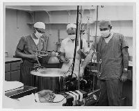
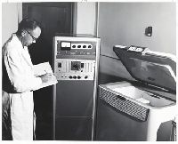
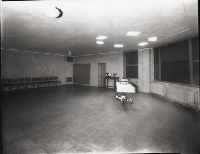
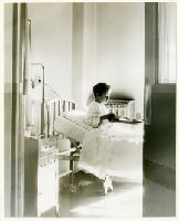
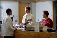
Attached statement: "An 'artificial kidney' used in illnesses when human kidneys are not functioning properly is shown in operation at the Yale-New Haven Medical Center. "Dr. Franklin H. Epstein (left) and Dr. Howard Levitin (right) together with nurse (center) operate the equipment connected by tubes to the artery of the patient's arm shown in foreground.... The 'artificial kidney' was purchased by a gift of $5000 to Yale from the United Fund of Middletown, Connecticut, March 1959"
Subject (Geographic):
United States
Subject (Name):
Epstein, Franklin H., 1924-2008, Levitin, Howard, 1928-2010, and Yale-New Haven Medical Center
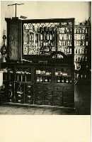
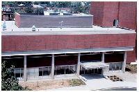
Inscription: Reproduced for Associates of the Yale Medical Library; Printed by The Meriden Gravure Company, Meriden, Conn. and The pharmacy was removed from the lobby outside the medical library at the time of a major library renovation ca. 1989.
Publisher:
Associates of Yale Medical Library
Subject (Name):
Streeter, Edward Clark, 1874-1947
Subject (Topic):
Medical libraries, New Haven (Conn.), Pharmacies, and Yale Medical Library
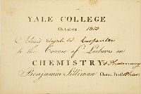
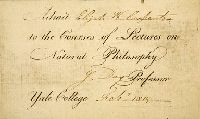
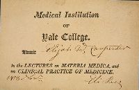
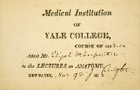
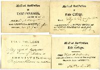
Four engraved admission cards for Elijah W. carpenter of Vermont to attend the first set of lectures given at the Medical Institution of Yale College in 1813. The cards are signed by Nathan Smith, Eli Ives, Jonathan Knight, and Benjamin Silliman. Carpenter attended the medical school for one year and did not receive an M.D. (which was not required to practice medicine). The card are part of the Elijah W. Carpenter papers.
Subject (Geographic):
United States
Subject (Name):
Carpenter, Elijah W., 1778-1855, Ives, Eli, 1779-1861, Knight, J. (Jonathan), 1789-1864, Silliman, Benjamin, 1779-1864, Smith, Nathan, 1762-1829, and Yale University. School of Medicine
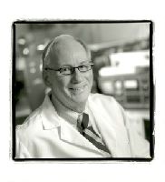
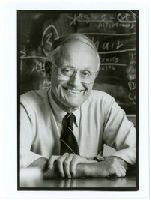
DuBois, A. B. (Arthur Brooks), Yale University. School of Medicine. Department of Cellular and Molecular Physiology, and Yale University. School of Medicine. School of Epidemiology and Public Health
Subject (Topic):
Epidemiologists, Faculty, Medical, Laboratories, and Physiologists
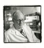
DuBois, A. B. (Arthur Brooks), Yale University. School of Medicine. Department of Cellular and Molecular Physiology, and Yale University. School of Medicine. School of Epidemiology and Public Health
Subject (Topic):
Epidemiologists, Faculty, Medical, Laboratories, and Physiologists
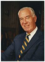
Ebbert, Arthur, Jr., 1922-2012, Yale University. School of Medicine. Department of Internal Medicine, and Yale University. School of Medicine--Administration
Subject (Topic):
Deans (Education), Faculty, Medical, and Internists
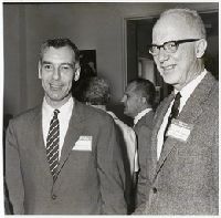
Photograph taken on Alumni Day, May 24, 1969. Nicholas Spinelli is in the background.
Subject (Geographic):
United States
Subject (Name):
Ebbert, Arthur, Jr., 1922-2012, Forbes, Thomas Rogers, 1911-1988, Spinelli, Nicholas, 1921-2007, and Yale University. School of Medicine--Administration
Ebbert, Arthur, Jr., 1922-2012, Lippard, Vernon W., 1905-1985, Yale University. School of Medicine. Department of Internal Medicine, Yale University. School of Medicine. Department of Pediatrics, and Yale University. School of Medicine--Administration
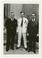
Drs. Yudkin and Blake were clinical professors of ophthalmology. Smith is unidentified. Photograph was taken outside Sterling Hall of Medicine.
Subject (Geographic):
United States
Subject (Name):
Blake, Eugene Maurice, 1882-1969, Smith, Byron C., 1908-1990, Yale University. School of Medicine. Department of Ophthalmology, and Yudkin, Arthur Meyer, 1892-1967
Carl Van Vechten Papers Relating to African American Arts and Letters
Container / Volume:
Box 111 | Folder 2077
Image Count:
2
Resource Type:
Prints & Photographs
Description:
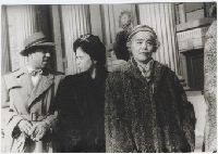
Snapshot taken outside of Yale Commons: Richmond Barthé, Ellen Tarry, Dorothy Peterson. and Stamped verso: "Photograph by Carl Van Vechten. / 101 Central Park West / Cannot be reproduced without permission.
Subject (Name):
Barthe, Richmond, 1901-1989, Peterson, Dorothy, and Tarry, Ellen, 1906-
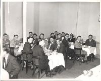
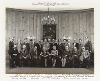
Beaumont Room, Yale University School of Medicine, October 7, 1977. Back row: L. H. Wardner, John D. Thompson, D. Crombie, R. S. Beckett, J. A. Kirchner, A. Bouhuys, Sherwin B. Nuland (President), Thomas R. Forbes, Arthur Viseltear (Secretary), W. G. Downs, Dorothy Horstmann, Ferenc Gyorgyey, Martin E. Gordon, N. Canfield, Alvan R. Feinstein, H. Mark. 1st Row: A. S. Evans, J. W. Meigs, A. F. Miller, Madeline E. Stanton, Elizabeth A. Thompson, Lucia Fulton, William W. L. Glenn, Ulrich H. Weil.
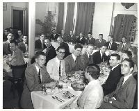
Front row: Field, Bill (William) 1917-1993; Horstsman, Dorothy Millicent 1911-2001; Musto, David 1936-2010; Holmes, Frederick (Larry) 1937-2003; Leonard Wilson. Back row: Viseltear, Arthur J 1938-1990; Spiro, Howard Marget 1924-2012; Stevenson, Lloyd 1918-1988; Bylebyl, Jerome 1946-2008; Bob Maury. Actual portrait can be found in the Beaumont Room.
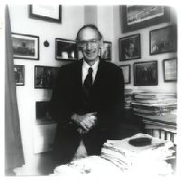
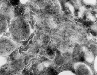
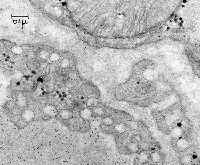
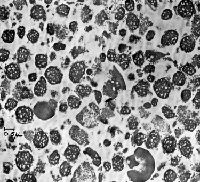
3.25 in. x 4 in. Lantern Slides, Original Magnification: x20,000, and This was the first study to use immuno-EM to localize secretory proteins in pancreatic acinar cells. It showed that the RER cisternae, all compartments of the Golgi and all zymogen granules contained trypsinogen. We also demonstrated that trypsinogen was much more concentrated in zymogen granules than in the earlier compartments on the secretory pathway.
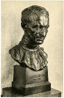
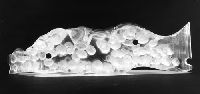
Inscription: Reproduced for Associates of the Yale Medical Library; Printed by The Meriden Gravure Company, Meriden, Conn. and The bust of Cushing is in the Reading Room of the Historical Library.
Publisher:
Associates of Yale Medical Library
Subject (Name):
Cushing, Harvey, 1869-1939 and Hoffman, Malvina, 1887-1966
Subject (Topic):
Medical libraries, New Haven (Conn.), Portrait sculpture, and Yale Medical Library
Palade, George E Simeonescu, Maya Simeonescu, Nicolae
Published / Created:
10/24/1961
Image Count:
1
Resource Type:
Prints & Photographs
Description:
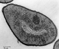
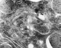
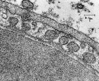
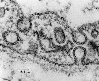
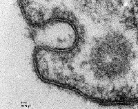
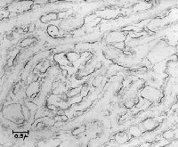
3.25 in. x 4 in. Lantern Slides, Hemoglobin, used as a tracer, is seen in the lumen of a capillary lined by a continuous endothelium, in endothelial caveolae, and in the extracellular space. Tracers such as hemoglobin or small heme-peptides were used by these investigators to demonstrate the role of caveolae in transcytosis of molecules across continuous capillary endothelial cells., and Original Magnification: x84,000
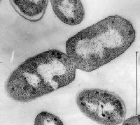
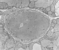
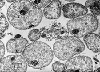
3.25 in. x 4 in. Lantern Slides, Original Magnification: x18,000, and Rat diaphragm skeletal muscle reticulocyte in capillary lumen; continuous endothelium.
3.25 in. x 4 in. Lantern Slides, One of Dr. Palade's long standing interests concerned the structure and function of capillaries. He and his many colleagues identified a variety of types of capillary endothelium (fenestrated, continuous) and determined the role of caveolae in transporting macromolecules across endothelial cells. Collaborators included: Nicolae and Maya Simeonescu and Romaine Bruns., and Original Magnification: x88,000
3.25 in. x 4 in. Lantern Slides, Clathrin coated pit caveola and transgential cut of coated vesicle in endothelial cell., and Original Magnification: x248,000
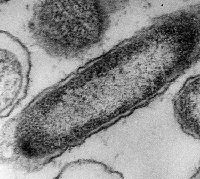
3.25 in. x 4 in. Lantern Slides, Discontinuous capillary; guinea pig pancreas; pericyte under the endothelium. Red blood cell in lumen., and Original Magnification: x17,000
3.25 in. x 4 in. Lantern Slides, Capillary lumen rat diaphragm 24 hr. post IV ferritin. Some ferritin in intracellular vesicles and extracellular space., and Original Magnification: x25,000
3.25 in. x 4 in. Lantern Slides, Capillary rat diaphragm 2h post IV ferritin. Ferritin is seen entering plasmalemmal vesicles and into the extracellular space., and Original Magnification: x62,000
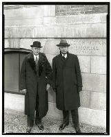
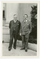
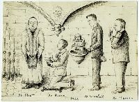
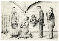
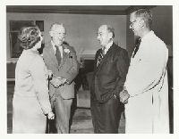
Dr. Morse is presumably showing Dr. Flint two offers, one from Iowa and one from Yale.
Subject (Geographic):
United States
Subject (Name):
Creadick, Abraham Nowell, 1883-1956, Flint, Joseph Marshall, 1872-1944, Harvey, Samuel Clark, 1886-1953, Morse, Arthur Henry, 1880-1950, and Yale University. School of Medicine. .
Filed in Joseph Marshall Flint folder. Another copy cataloged was from the Samuel C. Harvey folder. Shows Flint having to decide between competing offers to Morse from Iowa and Yale. Location of original drawing is unknown.
Subject (Geographic):
United States
Subject (Name):
Creadick, Abraham Nowell, 1883-1956, Flint, Joseph Marshall, 1872-1944, Harvey, Samuel Clark, 1886-1953, Morse, Arthur Henry, 1880-1950, Yale University. School of Medicine. Department of Obstetrics a, and Yale University. School of Medicine. Department of Surgery
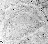
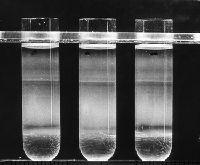
3.25 in. x 4 in. Lantern Slides, Original Magnification: x13,750, and The authors took advantage of the fact that rats gavage-fed a large bolus of ethanol (single malt I hope) were observed to have their Golgi cisternae loaded with LDL particles. Since LDL particles are lighter than most other intracellular organelles, following homogenization of ethanol-fed rats, the Golgi fractions "floated" to the top of denser sucrose gradients during centrifugation. This allowed investigators to analyze the membrane properties of "pure" Golgi fractions and set the stage for development of fusion assays that led to an understanding of factors regulating the specificity of membrane fusion - the SNARE hypothesis.
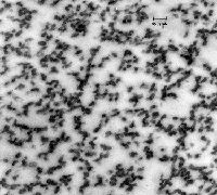
3.25 in. x 4 in. Lantern Slides, Original Magnification: x54,000, and Rough microsomes, the cell fractionation equivalent of the rough ER in intact cells, were extensively used to determine the mechanism of translocation of newly synthesized exportable proteins across the ER membrane. The culmination of these studies on rough microsomes was the elucidation of the "Signal Hypothesis".
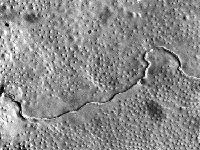
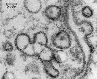
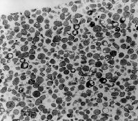
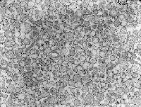
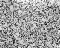
3.25 in. x 4 in. Lantern Slides, Cell fractionation confirmed that newly synthesized acinar cell secretory proteins moved from the RER to the periphery of the Golgi in the lumen of a smooth-surfaced membrane bounded compartment. This paper also describes the pancreatic slice system and the pulse-chase protocol used to label newly synthesized secretory proteins with radioactive leucine. The micrograph shows smooth microsomes obtained from the top band in the photograph of sucrose density gradient., and Original Magnification: x10,000
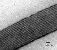
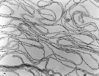
3.25 in. x 4 in. Lantern Slides, Dr. V. T. Marchesi from Yale and colleagues took advantage of the high purity of red cell ghosts obtained by hypotonic lysis to analyze the properties of membrane proteins. They identified spectrin (from the Latin specere, to look at - or ghost) as a major red cell membrane cytoskeletal protein responsible for maintaining the biconcave shape of red cells. In hereditary spherocytosis, spectrin is mutated resulting in anemias due to greater fragility of red cells which cannot assume a biconcave shape., and Original Magnification: x25,000
3.25 in. x 4 in. Lantern Slides and Cell fractionation confirmed that newly synthesized pancreatic acinar cell secretory proteins moved from the RER to the periphery of the Golgi in the lumen of a smooth-surfaced membrane bounded compartment. While EM autoradiography told us a great deal about the route and timetable of intracellular transport of nascent proteins, limitations of resolution of the technique (~0.1 micron) precluded a definitive localization to a cell compartment. This was provided by cell fractionation in which rough and smooth microsomes could be obtained in relative purity from pulse labeled pancreatic slices and further fractionated into lumenal content of the microsomes and membranes. The data clearly showed that nascent proteins were always in transit through the cell in the lumen of vesicles derived from the RER and Golgi complex, were not associated with the membranes, and did not travel in soluble form through the cytosol. The top band in this linear sucrose gradient (density of 1.14 to 1.27 gm/cc) contained primarily smooth/Golgi microsome while the largest band in the bottom half of the gradient consisted of relatively pure rough microsomes.
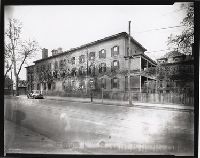
The Connecticut Training School for Nurses, Class of 1899. From left to right: Ann Clark, Lottie Bickford, Susan Burr, Grace Warrington, Mary McDermott, Harriet Benedict, Augusta Hickman, Isabel Garner.