
You Searched For
1 - 4 of 4
Search Results
- Creator:
- Jamieson, James D
- Published / Created:
- 9/8/1964
- Image Count:
- 1
- Resource Type:
- Prints & Photographs
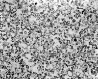
- Description:
- 3.25 in. x 4 in. Lantern Slides and Original Magnification: x10,000
- Publisher:
- Yale University School of Medicine
- Subject (Name):
- Jamieson, James D
- Subject (Topic):
- Cell Fractionation
- Found in:
- Medical Historical Library, Cushing/Whitney Medical Library > Cell Fractionation rough microsome fraction guinea pig pancreas
- Creator:
- Jamieson, James D
- Published / Created:
- 5/1/1961
- Image Count:
- 1
- Resource Type:
- Prints & Photographs
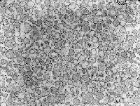
- Description:
- 3.25 in. x 4 in. Lantern Slides, Cell fractionation confirmed that newly synthesized acinar cell secretory proteins moved from the RER to the periphery of the Golgi in the lumen of a smooth-surfaced membrane bounded compartment. This paper also describes the pancreatic slice system and the pulse-chase protocol used to label newly synthesized secretory proteins with radioactive leucine. The micrograph shows smooth microsomes obtained from the top band in the photograph of sucrose density gradient., and Original Magnification: x10,000
- Publisher:
- Yale University School of Medicine
- Subject (Name):
- Jamieson, James D
- Subject (Topic):
- Cell Fractionation
- Found in:
- Medical Historical Library, Cushing/Whitney Medical Library > Cell Fractionation smooth microsome fraction guinea pig pancreas
- Creator:
- Jamieson, James D
- Published / Created:
- 3/10/1960
- Image Count:
- 1
- Resource Type:
- Prints & Photographs
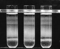
- Description:
- 3.25 in. x 4 in. Lantern Slides and Cell fractionation confirmed that newly synthesized pancreatic acinar cell secretory proteins moved from the RER to the periphery of the Golgi in the lumen of a smooth-surfaced membrane bounded compartment. While EM autoradiography told us a great deal about the route and timetable of intracellular transport of nascent proteins, limitations of resolution of the technique (~0.1 micron) precluded a definitive localization to a cell compartment. This was provided by cell fractionation in which rough and smooth microsomes could be obtained in relative purity from pulse labeled pancreatic slices and further fractionated into lumenal content of the microsomes and membranes. The data clearly showed that nascent proteins were always in transit through the cell in the lumen of vesicles derived from the RER and Golgi complex, were not associated with the membranes, and did not travel in soluble form through the cytosol. The top band in this linear sucrose gradient (density of 1.14 to 1.27 gm/cc) contained primarily smooth/Golgi microsome while the largest band in the bottom half of the gradient consisted of relatively pure rough microsomes.
- Publisher:
- Yale University School of Medicine
- Subject (Name):
- Jamieson, James D
- Subject (Topic):
- Cell Fractionation
- Found in:
- Medical Historical Library, Cushing/Whitney Medical Library > Cell Fractionation: sucrose density gradient separation of guinea pig pancreas microsomes