Palade, George E Simeonescu, Maya Simeonescu, Nicolae
Published / Created:
10/24/1961
Image Count:
1
Resource Type:
Prints & Photographs
Description:
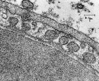
3.25 in. x 4 in. Lantern Slides, Hemoglobin, used as a tracer, is seen in the lumen of a capillary lined by a continuous endothelium, in endothelial caveolae, and in the extracellular space. Tracers such as hemoglobin or small heme-peptides were used by these investigators to demonstrate the role of caveolae in transcytosis of molecules across continuous capillary endothelial cells., and Original Magnification: x84,000
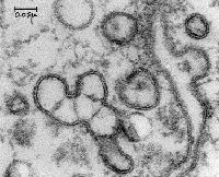
3.25 in. x 4 in. Lantern Slides, Original Magnification: x18,000, and Rat diaphragm skeletal muscle reticulocyte in capillary lumen; continuous endothelium.
3.25 in. x 4 in. Lantern Slides, One of Dr. Palade's long standing interests concerned the structure and function of capillaries. He and his many colleagues identified a variety of types of capillary endothelium (fenestrated, continuous) and determined the role of caveolae in transporting macromolecules across endothelial cells. Collaborators included: Nicolae and Maya Simeonescu and Romaine Bruns., and Original Magnification: x88,000
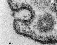
3.25 in. x 4 in. Lantern Slides, Clathrin coated pit caveola and transgential cut of coated vesicle in endothelial cell., and Original Magnification: x248,000
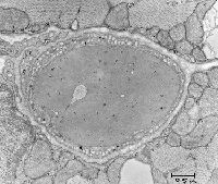
3.25 in. x 4 in. Lantern Slides, Discontinuous capillary; guinea pig pancreas; pericyte under the endothelium. Red blood cell in lumen., and Original Magnification: x17,000
3.25 in. x 4 in. Lantern Slides, Original Magnification: x13,750, and The authors took advantage of the fact that rats gavage-fed a large bolus of ethanol (single malt I hope) were observed to have their Golgi cisternae loaded with LDL particles. Since LDL particles are lighter than most other intracellular organelles, following homogenization of ethanol-fed rats, the Golgi fractions "floated" to the top of denser sucrose gradients during centrifugation. This allowed investigators to analyze the membrane properties of "pure" Golgi fractions and set the stage for development of fusion assays that led to an understanding of factors regulating the specificity of membrane fusion - the SNARE hypothesis.