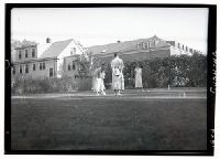
When the Cushing family arrived in the Boston area in 1912, the hospital building still wasn't ready for occupation. On the occasion of a visit by William Osler to Boston, an unofficial opening took place in February, 1913, the event captured in this phot
Subject (Name):
Cannon, Walter B. (Walter Bradford), 1871-1945, Christian, Henry Asbury, 1876-1951, Councilman, W. T. (William Thomas), 1854-1933, Cushing, Harvey, 1869-1939, Osler, William, Sir, 1849-1919, Peter Bent Brigham Hospital, and Warren, John Collins, 1842-1927
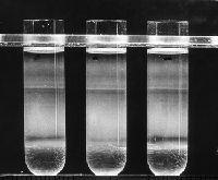
3.25 in. x 4 in. Lantern Slides and Cell fractionation confirmed that newly synthesized pancreatic acinar cell secretory proteins moved from the RER to the periphery of the Golgi in the lumen of a smooth-surfaced membrane bounded compartment. While EM autoradiography told us a great deal about the route and timetable of intracellular transport of nascent proteins, limitations of resolution of the technique (~0.1 micron) precluded a definitive localization to a cell compartment. This was provided by cell fractionation in which rough and smooth microsomes could be obtained in relative purity from pulse labeled pancreatic slices and further fractionated into lumenal content of the microsomes and membranes. The data clearly showed that nascent proteins were always in transit through the cell in the lumen of vesicles derived from the RER and Golgi complex, were not associated with the membranes, and did not travel in soluble form through the cytosol. The top band in this linear sucrose gradient (density of 1.14 to 1.27 gm/cc) contained primarily smooth/Golgi microsome while the largest band in the bottom half of the gradient consisted of relatively pure rough microsomes.